Digital Breast Tomosynthesis
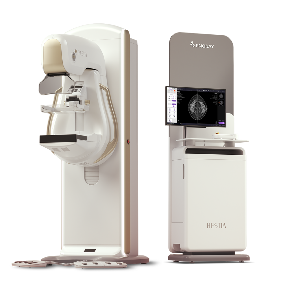
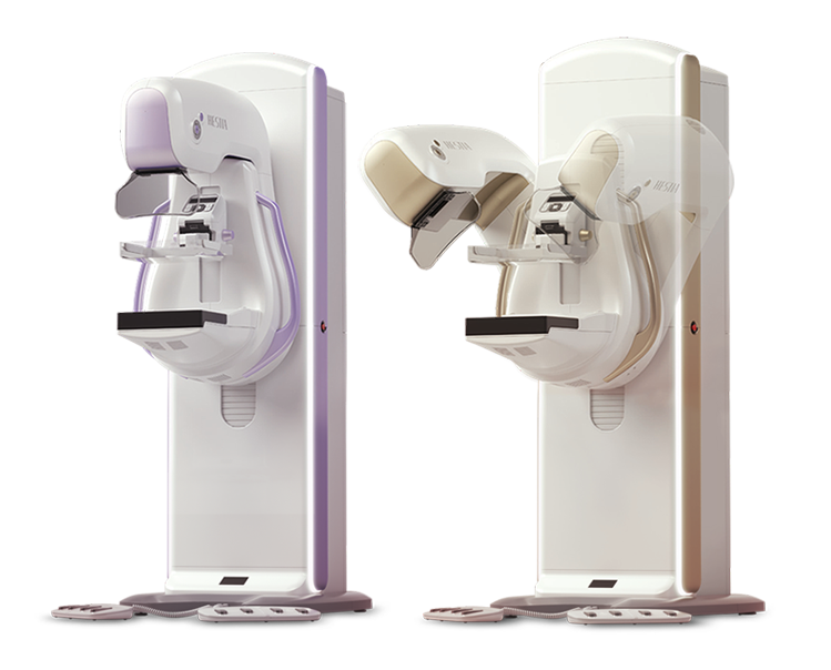
- Tomosynthesis(3D) Mammography System
- Tomosynthesis(3D) Mammography System
- Iso-centric Gantry
- Tomosynthesis FPD 24x30cm(TFT)
- 0kW HFG
- Tungsten(W) Target
- Low Dose & High-Quality Image
- Wide SID (660mm)
- Automatic Sequential Positioning System
DBT (Digital Breast Tomosynthesis)
The X-ray tube rotates around the breast and takes high-definition, high-resolution images with high contrast from various angles.
The images are reconstructed in 3D to provide accurate information about the location and condition of the lesion.

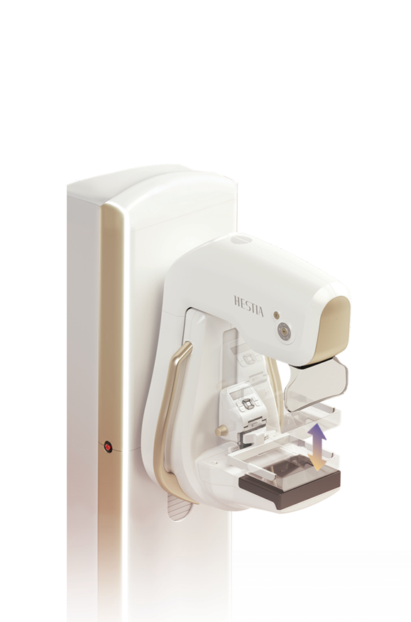
Synthesized 2D
HESTIA supports “Synthetic 2D” that generates 2D images only by tomography shooting.
VOICE / VISUAL GUIDE
HESTIA tells Examinee the current status of diagnosis by voice guide and instruction screen.
Automatic Compression-release Management Function
To ensure patient safety, HESTIA provides and manages an automatic compression-release function.
LED STATUS INDICATOR
HESTIA’S LED light gives its status in color to the operational real time.
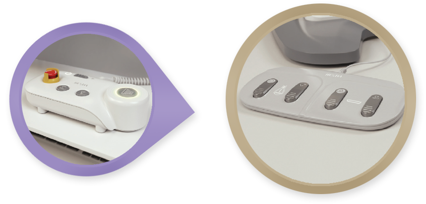
USER FRIENDLY OPERA
For the examine’s safety, there is “Emergency Stop Switch” on the operation pane.l
4-BUTTON FOOT SWITCH
HESITA is designed to allow for intuitive breast compression and up-and-down movement of the gantry.
UPGRADEABLE
UPGRADING FROM 2D to Tomosynthesis
The L (Large) type that could not only take 2D pictures can be upgraded into the T (Tomo) type that can also take 3D shots, it can be done with a simple software installation witoout additional H/W installation or system replacement.
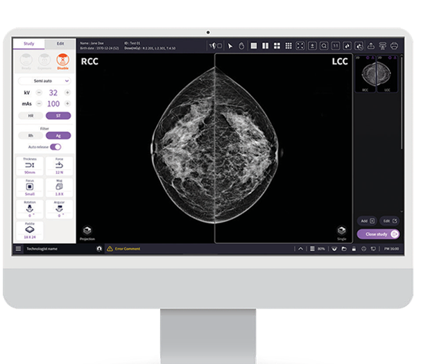
DEDICATED SOFTWARE
Dedicated Console Software “VENUS”
Viewer optimized for Mammography’s clinical workflow.
Optimized image Acquisition & processing Workstation for HESTIA.
Supports DICOM 3.0 and realizes PACS linkage.
For the operator’s convenience, HESTIA adopted Intuitive, highly visible user interface.